Symptoms of DVT in the leg are:
- Throbbing or cramping pain in 1 leg (rarely both legs), usually in the calf or thigh
- Swelling in 1 leg (rarely both legs)
- Warm skin around the painful area
- Red or darkened skin around the painful area
- Swollen veins that are hard or sore when you touch them
Once you have been referred to Diagnostic Healthcare for your DVT ultrasound scan, your referral letter will be processed immediately by our dedicated DVT team who will be in contact with you.
What is an ultrasound scan?
Ultrasound is a very safe and well tolerated procedure that uses high-frequency sound waves to create an image of part of the inside of the body. It is used to detect changes in the look, size or outline of organs, tissues and vessels, or to detect abnormal masses.It is a painless examination with no known harmful effects to humans.
What preparation is required for a DVT ultrasound?
A DVT scan requires no special preparation.What do we need to know before the scan?
If you are allergic to latex, it is important to inform our ultrasound team. In these cases, the team will use latex-free materials.What happens at the appointment and during an ultrasound scan?
Ultrasound examinations are usually painless and easily tolerated by most patients. Most types of ultrasound examinations are completed within 15 to 30 minutes and are performed by either a Radiologist or a Sonographer.When you arrive at our clinic for your appointment, our Sonographer/Radiologist and the Diagnostic Imaging Assistant will provide you with a full briefing on what will happen during your appointment.
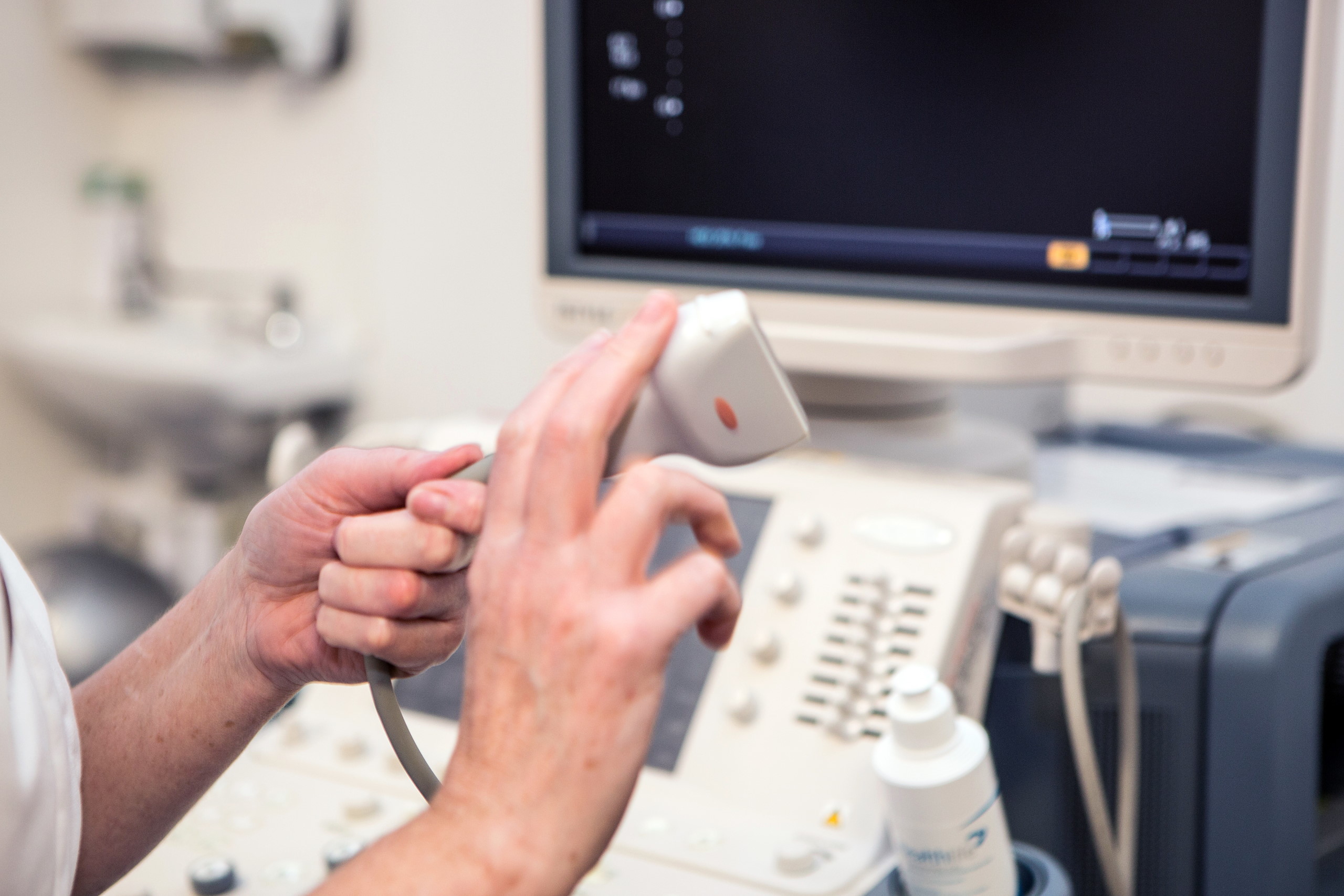
In most cases, you will lie on a medical couch and the clinician will apply a water-based gel on the area to be scanned and then pass a small handheld probe over the skin and over the part of the body being examined.
After the ultrasound scan
After the ultrasound scan has been completed, the images will be reviewed by the Sonographer/Radiologist who performed the scan and a report will be produced and sent to your doctor on the same day.There are no restrictions on normal activity, you can eat and drink normally, drive and return to work immediately after the scan.
Follow-up examinations may be necessary and recommendations may be included in your report. Sometimes a follow-up exam is done because a suspicious or questionable finding needs clarification with additional views or a special imaging technique.
A follow-up examination may also be necessary so that any change in a known abnormality can be monitored over time. Follow-up examinations are sometimes the best way to see if treatment is working or if an abnormality is stable over time. The report will be sent to your doctor.
Ultrasound FAQs
The ultrasound scan takes 15 to 30 minutes during which time you may be asked to change position to allow the area to be examined from different angles.
Ultrasound scans are a simple examination; they are non-invasive and are not painful. There may be varying degrees of discomfort from pressure as the transducer is pressed against the area being examined.
For standard diagnostic ultrasound there are no known harmful effects on humans.
The ultrasound scanner consists of a console (computer), a video display screen and a transducer (probe) that is used to scan the body.
The ultrasound scan report will be sent to your GP or referring clinician usually within 24-48 hours of the examination.


